Anatomy Of Chest Wall / The chest wall is formed from the sternum anteriorly, 12 pairs of ribs, costal cartilages and intercostal muscles laterally, and the thoracic vertebrae posteriorly.
Anatomy Of Chest Wall / The chest wall is formed from the sternum anteriorly, 12 pairs of ribs, costal cartilages and intercostal muscles laterally, and the thoracic vertebrae posteriorly.. Chest wall anatomy the chest is considered to be the area between the neck and the abdomen and contains many major organs as well as muscle groups. The palpable midline sternum is variable in size and shape; Describe the structure of the chest wall and diaphragm and to relate these to respiratory mechanics. 4 innervation of the breast blood supply of the breast syllabus p. The first rib is a short, flat rib that is much wider and more curved than those previously described.
Chest wall pain is caused by problems affecting the muscles, bones and/or nerves of the chest wall. Pectus excavatum is a congenital deformity of the ribs and the sternum producing a concave appearance of the anterior chest wall. The chest wall has 10 layers, namely (from superficial to deep) skin (epidermis and dermis), superficial fascia, deep fascia and the invested extrinsic muscles (from the upper limbs), intrinsic muscles associated with the ribs (three layers of intercostal muscles), endothoracic fascia and parietal pleura. Doctors diagnose chest wall pain in at least 25% of patients who come to the emergency room for chest pain. 31 anatomy of the female breast syllabus p.
Chest Wall Amboss from media-us.amboss.com The chest wall is comprised of skin, fat, muscles, and the thoracic skeleton. Thoracic wall dissection anatomy description: The thorax is bound by bony structures including the 12 pairs of ribs and thoracic vertebrae, whilst also being supported by many ligaments and muscles. The chest wall has 10 layers, namely (from superficial to deep) skin (epidermis and dermis), superficial fascia, deep fascia and the invested extrinsic muscles (from the upper limbs), intrinsic muscles associated with the ribs (three layers of intercostal muscles), endothoracic fascia and parietal pleura. It is also detrimental in the movement of the upper arms. The first rib is a short, flat rib that is much wider and more curved than those previously described. 4 innervation of the breast blood supply of the breast syllabus p. Vena azygos lobe a common normal variant is the azygos lobe.
The chest wall is formed from the sternum anteriorly, 12 pairs of ribs, costal cartilages and intercostal muscles laterally, and the thoracic vertebrae posteriorly.
The palpable midline sternum is variable in size and shape; The muscles of the thorax are also important for the vital actions of. Chest wall involvement is an uncommon manifestation of tuberculosis (, 17) that may be due to contiguous spread from underlying pleural or pulmonary lesions, although hematogenous seeding without active pulmonary disease is more common. The chest or thorax is the region between the neck and diaphragm that encloses organs, such as the heart, lungs, esophagus, trachea, and thoracic diaphragm. Chest wall abscess and sinus tract formation occur in about 25% of cases (, 3). The chest wall functions as a protective cage around the vital organs of the body, and significant disruption of its structure can have dire respiratory and circulatory consequences. Principal functions are the protection of internal viscera and an expandable cylinder facilitating variable gas flow into the lungs. Vena azygos lobe a common normal variant is the azygos lobe. The chest wall has 10 layers, namely (from superficial to deep) skin (epidermis and dermis), superficial fascia, deep fascia and the invested extrinsic muscles (from the upper limbs), intrinsic muscles associated with the ribs (three layers of intercostal muscles), endothoracic fascia and parietal pleura. The chest wall is supplied by the posterior intercostal arteries arising from the aorta, the internal thoracic and the highest intercostals given off the subclavian artery, and the branches of the axillary artery (fig. The past several decades have seen a marked improvement in the management and reconstruction of complex chest wall de … Therefore in the dorsal and lateral chest the surgeon must proceed along the superior margin of the lower rib when puncturing in order to aspirate or to perform surgical procedures. Chest wall pain is caused by problems affecting the muscles, bones and/or nerves of the chest wall.
Chest wall anatomy the chest is considered to be the area between the neck and the abdomen and contains many major organs as well as muscle groups. It is made up of the manubrium superiorly, the body and the xiphisternum (figure 1). Use the mouse scroll wheel to move the images up and down alternatively use the tiny arrows (>>) on both side of the image to move the images.>>) on both side of the image to move the images. The chest wall protects the heart lungs and liver provides a flexible skeletal framework to stabilize the actions of the shoulder and arm and promotes respiratory movement all while reliably. The azygos lobe is created when a laterally displaced azygos vein makes a deep fissure in the upper part of the lung.
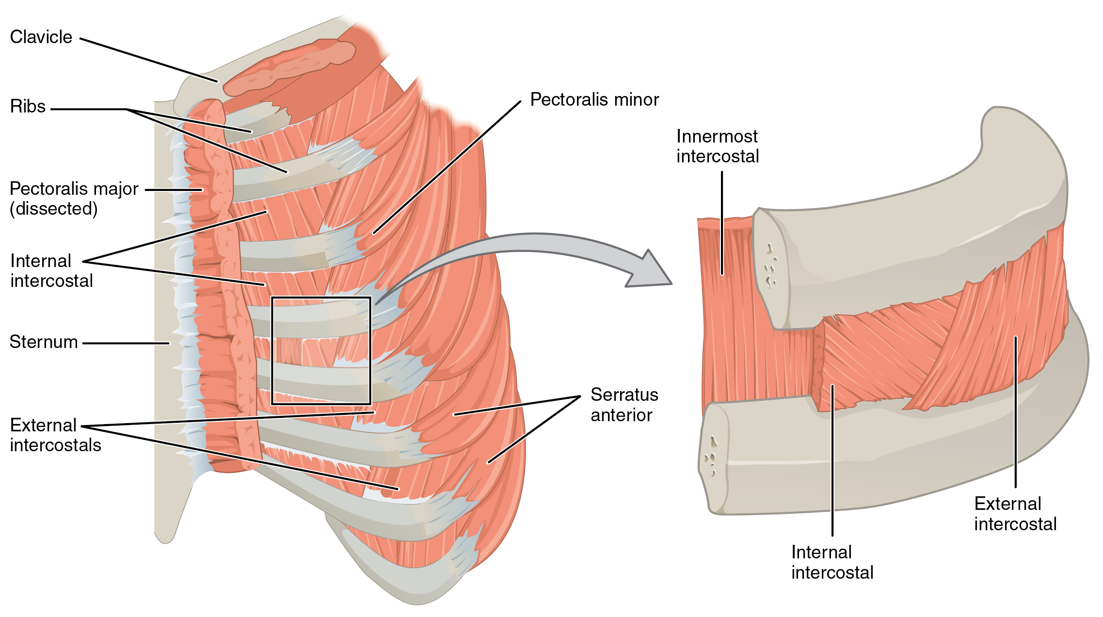
Figure 3 From Relevant Surgical Anatomy Of The Chest Wall Semantic Scholar from d3i71xaburhd42.cloudfront.net The chest wall has 10 layers, namely (from superficial to deep) skin (epidermis and dermis), superficial fascia, deep fascia and the invested extrinsic muscles (from the upper limbs), intrinsic muscles associated with the ribs (three layers of intercostal muscles), endothoracic fascia and parietal pleura. The dominant muscle in the upper chest is the pectoralis major. Therefore in the dorsal and lateral chest the surgeon must proceed along the superior margin of the lower rib when puncturing in order to aspirate or to perform surgical procedures. The chest wall is formed from the sternum anteriorly, 12 pairs of ribs, costal cartilages and intercostal muscles laterally, and the thoracic vertebrae posteriorly. 31 anatomy of the female breast syllabus p. The thorax is bound by bony structures including the 12 pairs of ribs and thoracic vertebrae, whilst also being supported by many ligaments and muscles. The thorax itself can be split up into various areas that contain important structures. Anatomy and the physiology of the chest wall ad sternum with surgical implications.
Use the mouse scroll wheel to move the images up and down alternatively use the tiny arrows (>>) on both side of the image to move the images.>>) on both side of the image to move the images.
Chest wall involvement is an uncommon manifestation of tuberculosis (, 17) that may be due to contiguous spread from underlying pleural or pulmonary lesions, although hematogenous seeding without active pulmonary disease is more common. Anatomy of the chest wall. 4 innervation of the breast blood supply of the breast syllabus p. Anterior chest wall showing muscular attachments and neurovascular structures ribs 3 through 9 are typical ribs as described earlier while ribs 1, 2, 10, 11, and 12 are atypical. Chest wall anatomy the chest is considered to be the area between the neck and the abdomen and contains many major organs as well as muscle groups. 2 skin of the anterior chest wall syllabus p. 30 lines of the thoracic wall syllabus p. 31 anatomy of the female breast syllabus p. Anatomy and the physiology of the chest wall ad sternum with surgical implications. The chest wall is what provides the protection needed for the vital organs such as the heart, lungs, liver and the major vessels. Chest wall abscess and sinus tract formation occur in about 25% of cases (, 3). Chest wall anatomy the chest is considered to be the area between the neck and the abdomen and contains many major organs as well as muscle groups. Chest wall pain is caused by problems affecting the muscles, bones and/or nerves of the chest wall.
The dominant muscle in the upper chest is the pectoralis major. The chest wall is what provides the protection needed for the vital organs such as the heart, lungs, liver and the major vessels. Use the mouse scroll wheel to move the images up and down alternatively use the tiny arrows (>>) on both side of the image to move the images.>>) on both side of the image to move the images. Pectus excavatum is a congenital deformity of the ribs and the sternum producing a concave appearance of the anterior chest wall. Vena azygos lobe a common normal variant is the azygos lobe.
Thoracic And Abdominal Muscles Lecturio Online Medical Library from d3uigcfkiiww0g.cloudfront.net The skeleton of the chest wall is composed of the sternum anteriorly and the spinal column posteriorly with twelve thoracic vertebrae and paired ribs (fig. The thoracic contents are bounded by the chest wall, providing both the shape of the thorax and protection for the intrathoracic contents ().the skin, subcutaneous tissues, and muscles that surround the rib cage and shoulder girdle appear radiographically indistinguishable from each other, whereas on ct the skin, fat, and muscles are recognized by their difference in attenuation. The normal chest wall is symmetric and broadens cranially to caudally. It contains skin, muscle and fatty tissues. The first rib is a short, flat rib that is much wider and more curved than those previously described. Describe the structure of the chest wall and diaphragm and to relate these to respiratory mechanics. The palpable midline sternum is variable in size and shape; Chest wall abscess and sinus tract formation occur in about 25% of cases (, 3).
The chest wall is formed from the sternum anteriorly, 12 pairs of ribs, costal cartilages and intercostal muscles laterally, and the thoracic vertebrae posteriorly.
30 lines of the thoracic wall syllabus p. The chest wall functions as a protective cage around the vital organs of the body, and significant disruption of its structure can have dire respiratory and circulatory consequences. The thorax is the area of the body situated between the neck and the abdomen. The muscles of the thorax are also important for the vital actions of. The mammary gland is located within the superficial fascia of the anterior thoracic wall. Unfortunately, in many cases, that's as far as the doctor takes the diagnosis. The chest wall is formed from the sternum anteriorly, 12 pairs of ribs, costal cartilages and intercostal muscles laterally, and the thoracic vertebrae posteriorly. The past several decades have seen a marked improvement in the management and reconstruction of complex chest wall de … Principal functions are the protection of internal viscera and an expandable cylinder facilitating variable gas flow into the lungs. Vena azygos lobe a common normal variant is the azygos lobe. 4 innervation of the breast blood supply of the breast syllabus p. This mri chest (thorax) axial cross sectional anatomy tool is absolutely free to use. Anatomy of the chest wall.
The thorax has two major openings: anatomy of chest. Chest wall involvement is an uncommon manifestation of tuberculosis (, 17) that may be due to contiguous spread from underlying pleural or pulmonary lesions, although hematogenous seeding without active pulmonary disease is more common.


Posting Komentar
0 Komentar